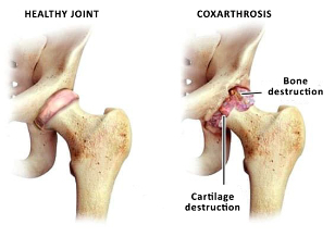
Osteoarthritis - a degenerative disease that leads to destruction of the hip joint and has a chronic nature of the flow. More common in older age groups. More common in women than men.
The beginning of the disease - a progressive, develops slowly. May affect one joint or both. Is the most common type of osteoarthritis.
Why develops the disease?
Osteoarthritis in some patients accompanied to the natural process of aging and Degeneration of the tissues of the hip joint is. Its appearance is influenced by such factors:
- reduced nutrition of the tissue;
- congenital malformation of the hip joint, in particular dysplasia;
- a Trauma suffered pelvic area;
- post coxarthrosis;
- aseptic osteonecrosis of the hip joint;
- Perthes disease (osteochondropathy).
Unfortunately, the cause of the disease determine is not always possible, and pathology of the hip joint is called idiopathic coxarthrosis, i.e. those whose causes are not installed. This is an incentive for the ongoing research problems. The scientific work in this area and the doctors came to the conclusion that a higher risk of coxarthrosis is for the following patients:
- An inherited tendency to pathology. Patients whose parents suffered from diseases of the cartilage and bone in most of the cases are also similar problems;
- Overweight. Significant body weight is the load on the joints, which is already regularly exposed to mechanical work;
- Metabolic diseases, Diabetes mellitus. This leads to a poor supply of oxygen and nutrients in the tissues of the joint, causing them to lose their properties.
Knowledge of the main risk factors of the disease, you can plan preventive measures to prevent its occurrence.
How to recognize the pathology of the hip joint?
Symptoms of osteoarthritis is dependent on the anatomical characteristics of the musculoskeletal system, the causes of the pathology and the stage of the process. You consider the most important clinical symptoms:
- Pain of the joint;
- radiating pain in the knee, hip, groin area;
- Stiffness of movement;
- limited mobility;
- Injury To The Foot, Lameness;
- reduced muscle mass of the thigh;
- Shortening of the injured limb.
The clinical picture corresponds to a set of internal changes in the tissues of the joint. Symptoms grow slowly and the first stages of the Patient does not pay them due attention. It is dangerous, because just at the beginning of the process of treatment brings about a greater effect.
Clinical and radiological degree of osteoarthritis
The following are the symptoms of the disease are characteristic for the respective degrees.
- 1 degree. The Patient will experience recurring pain and discomfort. Complaints bother after physical exertion, long-term Position in a static pose. Pain localized in the area of the joint and comes after the holiday. In this stage of the process, not pace and no shortening of the legs is disturbed. Changes in the x-ray image is visible - narrowed sustavnaya the gap, appear to be osteophytes (bony growths).
- 2 degrees. Increases the intensity of the pain you may be in the peace and radiates in the adjacent areas of the body. Appears lameness, after the person went long, or from over-exertion. The extent of movements in the joint is limited. Parallel to this, the radio develop logical changes in the work: the head of the femur, osteophytes grow on the inner and outer edges of the acetabulum.
- 3 stage. Pain of a permanent nature acquires, appears in the daytime and at night. Much worse gait, appears to be a permanent lameness. Strong motor skills, reduces the muscles of the legs atrophy. changing the muscle tissue causes the leg tightened a little and is shorter. This leads to the Deformation of the body posture and curvature of the body. X-rays in this Phase of the process: the total narrowing of the gap between the articular surfaces, the deformation of the femoral head, a significant growth of osteophytes.
Diagnostic program with the disease
The most important method of diagnosis is radiological. With its help, you the presence of the disease and its stage have to be determined. On radiographs, the structure of the joint of the subject matter of narrowing sustawnoj analyze Huckle the columns, osteophytes, destruction of the head.
If it is carried out is a necessity for the investigation of soft tissues - magnetic resonance imaging. It allows a detailed investigation of the condition of the cartilage phases of the joint, and the muscles of the hip.
Modern methods and directions of treatment of coxarthrosis of the hip joint
The treatment of osteoarthritis can be conservative and surgical. The treatment of coxarthrosis directed on achievement of the following objectives:
- To reduce the painful symptoms;
- Restoration of motor activity;
- Rehabilitation and restoration of ability to work;
- Prevention of complications;
- the improvement of the quality of life of the patient.
The beginning of the treatment consists in the modification of risk factors. For this doctor, the following measures are recommended:
- Normalization of body weight;
- The avoidance of harmful habits;
- Diet;
- Normalization of physical activity;
- balanced fluid intake;
- a healthy sleep.
Conservative treatment can be distinguished: - drug and non-drug. The drug treatment includes non-steroidal anti-inflammatory drugs, analgesics, chondro protectors. You can reduce to eliminate the inflammation in the tissues of the joint, swelling and pain, restore the mobility and improves the condition of the cartilage.
Drug-free treatment includes Massage of the affected area. It stimulates the muscles, contrary to their dystrophy, prevention, and shortening of the limb. A high-quality and professional Massage stimulates blood stream in the Zone of the joint, and this in turn leads to normalization of metabolism in the tissues. Please note that Massage is not always helpful, if coxarthrosis - it is only between exacerbations, and in some stages of the process. It is the treating physician can assign massage recommends techniques, the frequency rate of the procedure and the duration of the course.
A prerequisite of treatment - physiotherapy. Is the prevention of contractures and Progression of the disease. Exercises should be performed daily, only then you have effect. The gym is individual and from the doctor. The exercises improve the General health, reduces the risk for emotional disorders, strengthens the self-healing forces of the body.
Physiotherapy - a method of automatically when coxarthrosis. It may be, mud therapy, therapeutic baths and showers, magneto therapy. Hits electro - and phonophoresis of drugs.
If these treatment methods have not been brought to the effect or non - surgical treatment is required.
The surgical procedure in the case of coxarthrosis
The surgical treatment with the ineffectiveness of conservative methods. This is especially true in the case of later diagnosis. Modern surgical procedures and high-quality features operating system the structure and function of the joint, again, people, mobility and normal quality of life can restore. The most effective method of surgical treatment, arthroplasty, joint replacement is.
Indications for surgery are:
- coxarthrosis 2-3 degrees;
- the lack of effect of therapy;
- total restriction of movement to the foot.
Contraindications, do not allow the Operation:
- decompensated state of the kidneys, the heart, the liver;
- mental illness;
- the acute Phase of inflammation in the body.
This is due to the preoperative diagnosis. But if there is a way of correcting the condition - Patient is preparing for the surgery and after the Intervention takes place.
The Operation consists in the removal of the affected tissue, and the Installation of the prosthesis. There are different models of endoprostheses. The methods of their attachment to the bone – cement and cement, the Material from which the endoprosthesis is different. All the characteristics of the endoprosthesis and the intricacies of the Operation you get information on consultation with the attending physician.
Recovery period after surgery
From the first day after surgery, a Rehabilitation under the supervision of a physician is. First of all, it is the implementation of passive movements, then the load will be gradually increased. Walking in the first time is allowed only with crutches, can sitting and squatting.
Of course, in the first time after the Operation, there are restrictions on loads. Do not be afraid - because without surgery, these restrictions remained until the end of life. Decreased physical activity after surgery is necessary for the strengthening of the Position of the prosthesis, restoration of the integrity of the bones, wound healing. Within 2 months of sporting activities, physical loads on the joint, long Hiking and some types of exercises are excluded. After complete recovery, the person returns to normal life, can engage in sports and active sports.
The service life of the endoprosthesis: the majority of the companies is the survival rate of about 90% on the time limits for the monitoring of up to 15 years.


